What is Semen?
Semen is the combination of spermatozoa (sperm cells), and seminal fluid which is secreted by the reproductive glands of males. Seminal fluid provides a liquid medium through which sperm can move and it contains enzymes and fructose to promote sperm survival. Common presumptive screening tests for the presence of semen include the Acid Phosphatase Test and the Prostate Specific Antigen (p30) Test. These presumptive assays are only indicative of the presence of seminal fluid and are not dependent on the presence of sperm. Thus these tests will yield a positive result regardless of whether a male has been vasectomized or not.
Direct inspection by microscopy is necessary to confirm the presence of sperm. Sperm cells are typically the primary source of DNA for the individualization of a semen stain. Even in the case of vasectomized or otherwise aspermic males, however, it may be possible to obtain a DNA profile due to the presence of a small number of epithelial cells and leukocytes (white blood cells) in semen that can serve as a source of DNA.
What is the Acid Phosphatase Test? How specific is it and what can cause a false positive result?
Acid Phosphatase is an enzyme secreted by the prostate gland into seminal fluid. It is typically present at concentrations that are 20 to 400 times that of other body fluids. A purple color change with the addition of a few drops of sodium alpha naphthylphosphate and Fast Blue B solution indicates the presence of acid phosphatase. Large areas of fabric can be screened by pressing the material against a piece of moistened filter paper and then testing the filter paper for acid phosphatase activity. The presence of acid phosphatase serves only as a presumptive indication for the presence of semen and is not specific to humans. False acid-phosphatase positive results have been reported with a wide variety of materials including blood, vaginal discharge and some plant materials. Thus positive acid phosphase test results should be interpreted with great caution.
What is the Prostate Specific Antigen Test? How specific is it and what can cause a false positive result?
Prostate-specific antigen, also known as p30, is a protein produced by the prostate gland. The term “prostate-specific” antigen, however is a misnomer as it is not actually specific to the prostate. While it is mostly found in seminal fluid (300,000 – 5,500,000 ng/ml), it has also been detected (albeit in much lower quantities) in association with male and female urine, peripheral blood, some tumors, breast tissue, breast milk, the periurethral glands and amniotic fluid.
A p30-specific membrane test can be used for the forensic detection of semen. The underlying technology for these assay systems is analogous to that used with the HemDirect®/HemaTrace® assays for blood. These membrane tests are also used in clinical settings for the early detection of prostate cancer. Briefly, the test is based on an interaction between a monoclonal antibody targeted to human p30. These monoclonal antibodies are labeled with a pink dye. In the presence of human p30, the labeled monoclonal antibodies migrate across the test device and aggregate upon contact with a second, stationary phase antibody that also recognizes human p30. The aggregation of the labeled antibodies produces a pink line that is visible to the naked eye. The formation of this pink line serves as a presumptive indication of the presence of human p30 and thus by extension, human seminal fluid. The testing device also features an internal positive control that yields a second pink line indicating the test worked properly. Finally, the PSA Semiquant assay system also includes a 4 ng internal standard. This results in the formation of a third pink line that allows the analyst to estimate the concentration of p30 in a sample and thus to determine the how best to process the sample to obtain a DNA profile. Thus the presence of three pink lines indicates a positive result while the presence of only the control and internal standard lines indicates a negative result. See diagram below for image of mechanism.
The absence of either the control line or the internal standard line invalidates the test. These tests provide only a presumptive indication of human p30. This test is known to yield false positive results with the semen of higher-order primates. False indications of semen have also been reported in female urine as well as male urine and blood samples - especially from males with prostate cancer which contain elevated levels of p30.
What techniques are used for microscopic visualization of sperm?
|
Baecchi Stain, Christmas Tree Stain and Sperm HyLiter™ are the most common reagents employed for the staining and subsequent visualization of sperm. The visual identification of human spermatozoa (sperm cells) by microscopy is considered confirmatory for the presence of semen. It is important to recognize, however, that neither Baecchi Stain nor Christmas Tree Stain are species specific. Thus is necessary for a trained analyst to differentiate human from non-human sperm based on cell morphology and/or subtle differences in staining patterns. Artifacts such as yeast cells or other microbes may also complicate interpretation if they are incorrectly identified as sperm cells. This makes the accurate examination of fecal swabs or samples with significant microbial growth and/or decomposing sperm more challenging.
|
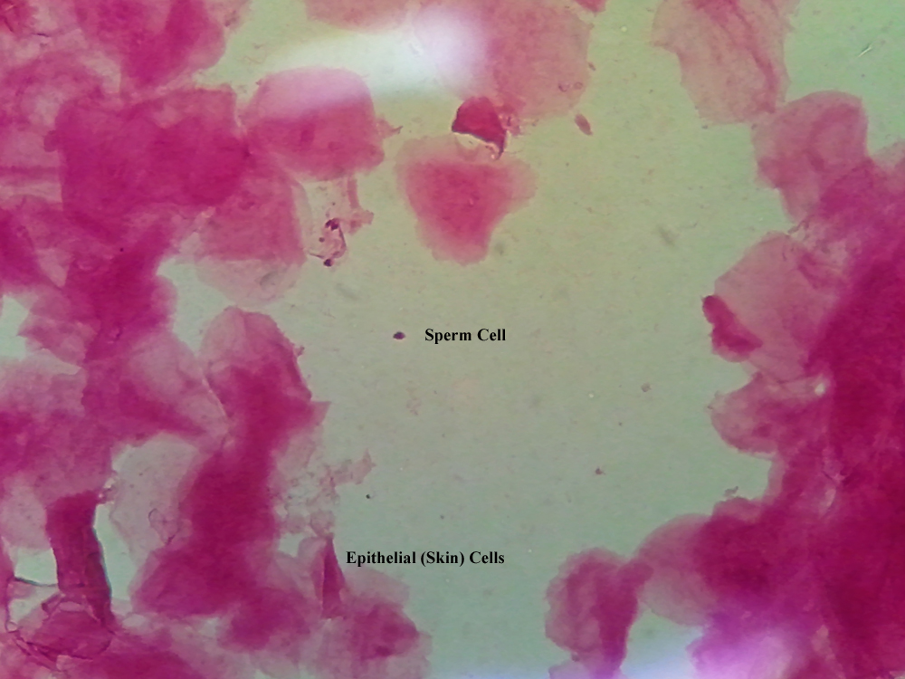
Baecchi Stain is a mixture of methylene blue and acid fuchsin. In the presence of this stain, the heads of sperm stain red, while the tails and mid-pieces stain blue. The cytoplasm of epithelial cells will stain pink while the nucleus will stain purple/blue. |
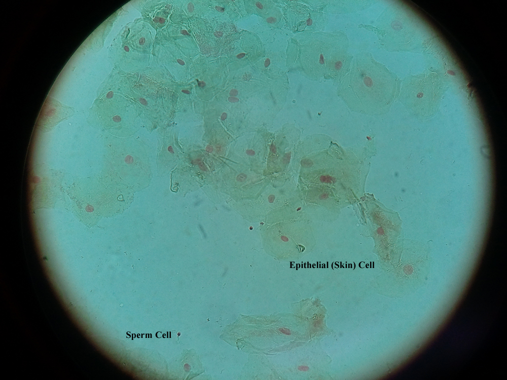
A common method of observing spermatozoa as part of a confirmatory testing is to recover dried semen evidence from fabric or human skin with a moistened swab. A portion of the recovered cells is then placed onto a microscope slide and heat-fixed to the slide. The immobilized cells are stained with Christmas Tree Stain which consists of aluminum sulfate, nuclear fast red, picric acid and indigo carmine. The stained slide is then examined under a light microscope for the presence of sperm cells. The Christmas Tree Stain marks the anterior of the sperm head light red / pink, the posterior of the head dark red, the sperm’s mid-piece blue, and the tail yellowish green. |
 |
 |
| |
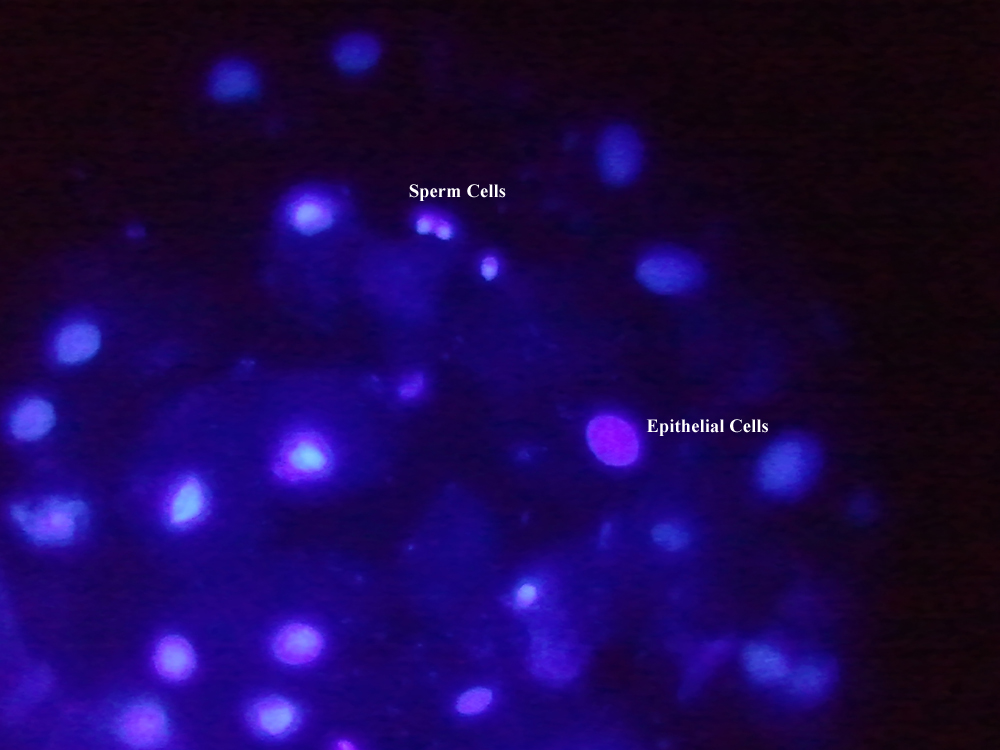
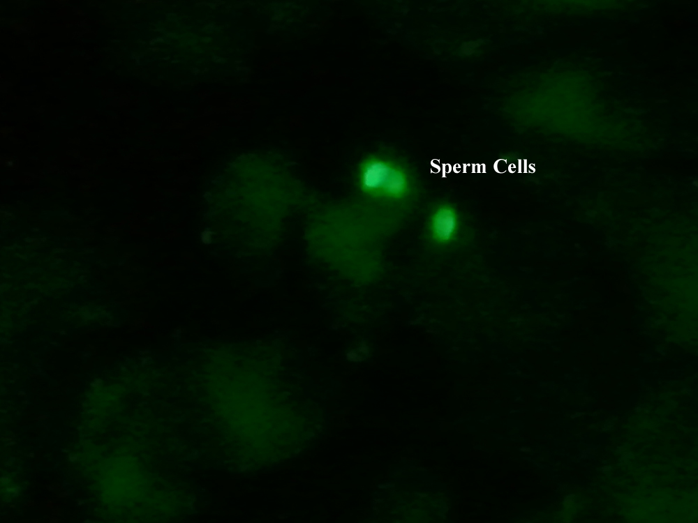
Sperm Hy-Liter™ uses a fluorescently tagged monoclonal antibody targeted to a human sperm head protein to detect the presence of human sperm cells. Sperm Hy-Liter™-stained slides must be visualized using a fluorescence microscope fitted with an appropriate filter set. The cell nuclei, of both epithelial and sperm cells appear as fluorescent blue bodies using DAPI-compatible filters while human sperm heads appear as fluorescent green bodies using fluorescein or Alexa 488 compatible filters. Sperm Hy-Liter™ is human-specific has not been reported to show cross-reactivity with non-human semen including that from horse, bull, sheep, goat, pig, dog, cat, mouse or primates. |
 |
 |
|